How is Gamma Knife Radiosurgery Performed?
You can watch the video above to see how Gamma Knife radiosurgery is performed.
Gamma Knife Radiosurgery
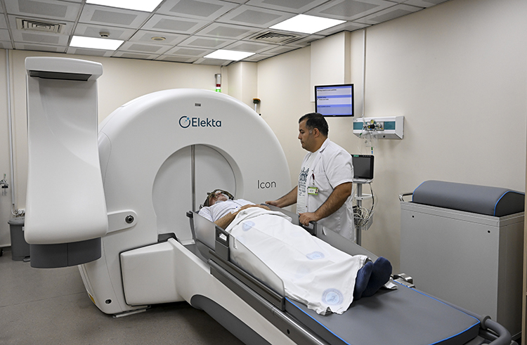
Gamma Knife technology was pioneered by Swedish neurosurgeon Lars Leksell. Following development in the 1950s, the first clinical treatments were performed in 1968. In Turkey, Gamma Knife treatment has been in clinical use since 1997. Since 2005, approximately 10,000 patients have been treated at Gazi University. In our clinic, treatment is performed using the modern Gamma Knife Icon platform.
What is Gamma Knife Radiosurgery?
Gamma Knife is not actually a knife. It is a radiosurgery method that uses sophisticated technology instead of a surgical blade, directing gamma rays with great precision to a target inside the skull in a single session. Like the precision expected from a scalpel, the aim is to treat the diseased area safely and effectively. Because no open surgery is performed, problems such as surgical bleeding, infection, and intensive care stay are avoided. With advanced imaging and planning techniques, very thin beams of gamma radiation can be directed to small areas inside the skull. Radiation beams from 201 gamma-ray sources focus on the diseased area, allowing the target tissue to receive a high dose while the surrounding normal brain tissue receives only a negligible dose. These steps are completed within a few hours, and the patient can usually return home on the same day.
Gamma Knife treatment is an outpatient procedure and consists of the following stages:
- Frame Installation
- Radiological Imaging
- Treatment Planning
- Treatment
TREATMENT STAGES
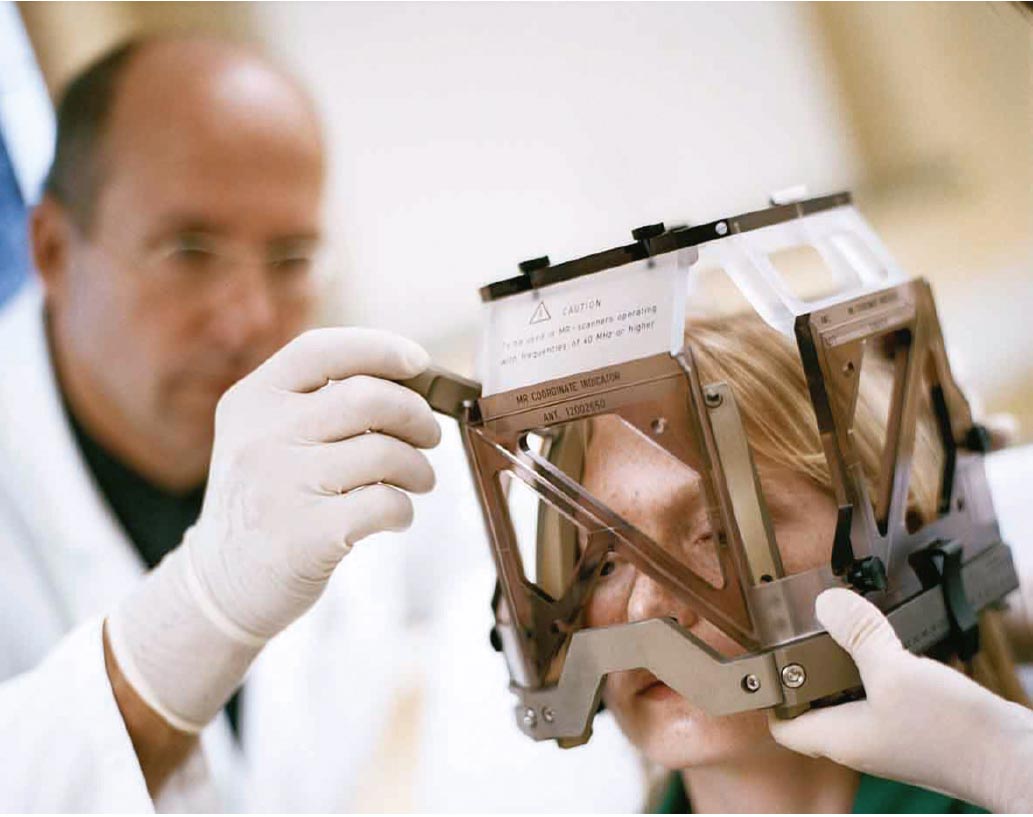
Frame: At the beginning of treatment, the Leksell stereotactic frame is attached to the patient's head. This is performed under local anesthesia. In patients younger than 10 years of age, the procedure is usually performed under sedation.
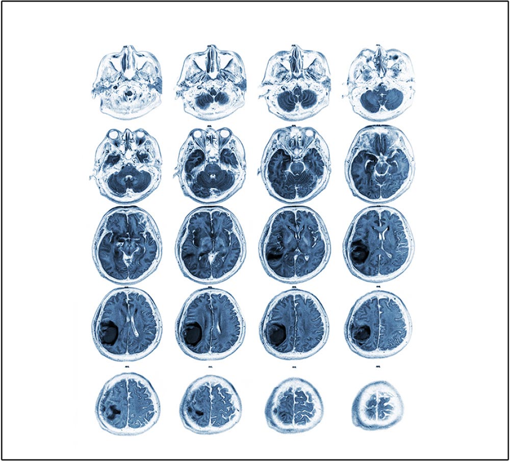
Imaging: After the frame is attached, imaging appropriate for the patient's condition is performed. Today, MRI is obtained in all patients. In AVM cases, angiography is performed with the stereotactic frame in place. In some skull base tumors, or in patients who cannot undergo MRI, CT may also be used for treatment planning.

Planning: The treatment target is contoured with high precision using dedicated planning software, and the radiation dose is calculated accordingly. For lesions near critical structures, selective beam-blocking ("plugging") can be used to prevent radiation from entering from specific directions. Modern planning software has significantly improved safety and patient comfort. During this stage, patients are typically awake and may talk or watch TV.
Treatment: After the treatment plan is prepared on the computer, the treatment is delivered in a separate treatment unit. This may take from 30 minutes to 3-4 hours. The main factors are the volume of the lesion and the radiation dose to be delivered.
Can you explain Gamma Knife treatment more simply?
Gamma Knife treatment is a high-dose RADIATION treatment. With advances in technology, this radiotherapy technique has become highly specialized: the normal parts of the brain receive almost no radiation, while only the targeted tissue, such as a tumor or vascular malformation, receives a high dose. This is the main advantage of Gamma Knife treatment and the reason it differs from many other radiotherapy applications. In this way, the normal structures of the brain are protected from the long-term effects of radiation as much as possible.
Is Gamma Knife treatment suitable for all types of brain disease?
Gamma Knife treatment is NOT suitable for every brain disease.
Gamma Knife treatment has many scientifically supported uses.
The most important include brain tumors, vascular malformations
of the brain, and some severe pain disorders.
However, Gamma Knife treatment cannot be used for every disease.
First, there should be scientific evidence that the condition to
be treated is likely to benefit from Gamma Knife treatment. In
addition, size is one of the most important criteria. The general
view is that Gamma Knife treatment is most suitable for brain
pathologies that are 3 cm or smaller.
With advances in device technology, and especially depending on the exact location of the disease within the brain, this size limit may be adjusted. On the other hand, as with any treatment, Gamma Knife treatment can have side effects. For example, if a tumor is very close to the optic nerves and Gamma Knife treatment may carry a long-term risk of vision loss, surgery may be preferred as the first option.


Is Gamma Knife treatment suitable for organs outside the brain?
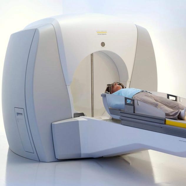
Gamma Knife treatment is a specialized method designed and used for diseases of the brain. With recent technological developments after the Gamma Knife Perfexion device, Gamma Knife radiosurgery can also treat selected conditions down to the level of the upper two cervical vertebrae. However, it cannot be used for tumors located lower in the body, such as liver or lung diseases. Although this may seem like a disadvantage at first, it is actually one of the device's most important features. Gamma Knife treatment draws its strength from being a Neurosurgery device. With technology that is not found in other devices today, it was developed specifically to treat brain diseases.
Are there any side effects of Gamma Knife treatment?
Almost every medical treatment involves some degree of risk, and every treatment also has a success rate. No treatment method is successful in 100% of cases. One of the most important features of Gamma Knife treatment is that these risks can often be reduced to very low levels. Depending on the type of disease and, especially, its location in the brain, high-dose radiation treatment may carry different risks. Compared with surgery, these risks are usually lower, but no treatment is completely risk-free. The best way to understand your individual risk profile is to discuss it in detail with your treating physician.
Will it hurt during Gamma Knife treatment?
Pain is not expected during Gamma Knife treatment. To perform the treatment, a special frame must be fixed to the skull at four points. This is done after the scalp is numbed with strong local anesthetic medication, and the discomfort is not usually more than what is felt during a blood draw. After the frame is placed, the remaining steps are imaging and treatment, and these are not painful. Because children may not be able to cooperate with frame placement, general anesthesia may be used when necessary, depending on the child's age, so that no discomfort is felt.
How long does Gamma Knife treatment last?
One of the most important advantages of Gamma Knife treatment is that it can be delivered as a same-day outpatient procedure. You can come to the clinic in the morning, complete the required steps, receive treatment, and return home in the evening. In patients who require general anesthesia, an overnight hospital stay may be recommended for safety.
Will Gamma Knife treatment be a definitive solution to my disease?
Gamma Knife has been used worldwide for more than 50 years and is
an effective treatment, but it is not a "magic wand." Expected
outcomes vary by diagnosis. For many brain tumors, success usually
means controlling growth (or reducing size), rather than completely
eliminating the lesion in every case. Because each pathology and
each patient's response is different, additional treatment may be
needed in selected cases.
In arteriovenous malformations (AVMs), the goal is complete closure
of the abnormal vessels. Gamma Knife achieves complete obliteration
in many patients (often around 70-80%, depending on AVM
characteristics). If the AVM is not fully closed after follow-up,
repeat radiosurgery or other treatment options may be considered.
I had Gamma Knife treatment for a disease in my brain. Do I still need to see a doctor?
Gamma Knife treatment is a form of radiotherapy, and the effect of radiation does not appear immediately. For example, it can take 2-3 years for a vascular malformation to close. Tumors also respond over time. For this reason, your follow-up with Neurosurgery does not end after treatment. Possible side effects, if they occur, may also appear over time rather than immediately after treatment. You should therefore attend regular check-ups and remain under your doctor's supervision.