Pediatric Neurosurgery · Information for Families
What Is Lipomyelomeningocele? Is Surgery Needed?
Lipomyelomeningocele is a congenital abnormality of spinal cord development. The child may appear completely normal at first; what is important is the preservation of the nerves controlling the legs, bladder, and intestines as the child grows.
Prof. Dr. Alp Özgün Börcek
Neurosurgery
Pediatric Neurosurgery
Ankara Güven Hastanesi
You can contact us to have your MRI images reviewed in conjunction with your previous test results.
Schedule an Appointment via WhatsApp Contact and examination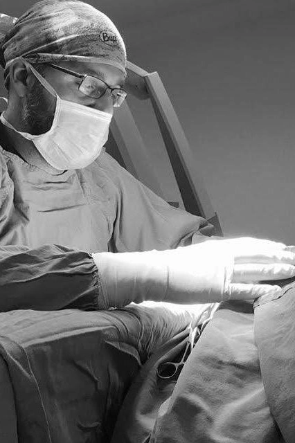
Prof. Dr. Alp Özgün Börcek
Neurosurgery
Pediatric Neurosurgery · Ankara Güven Hastanesi
This page has been prepared to help families better understand the topic. It is not intended as a diagnosis or treatment recommendation on its own; every child’s examination and MRI findings are unique.
What Is Lipomyelomeningocele?
Lipomyelomeningocele is a congenital condition in which the spinal cord tissue in the baby’s lower back is abnormally connected to fatty tissue. The main issue here is not the presence of fatty tissue itself, but rather that the spinal cord becomes trapped by this tissue and cannot move freely enough within the spinal canal.
As a child grows, the spine lengthens. If the spinal cord remains attached to surrounding tissues, it can become stretched over time. This condition is called a “tethered cord” and can affect the nerves that control the legs, bladder, or bowels.
The condition is present from birth; however, the problems it can cause sometimes don’t appear until months or years later.
How and When Does It Form?
The tissues that will form the baby’s brain and spinal cord begin to develop very early in pregnancy. In lipomyelomeningocele, a developmental abnormality occurs during the separation of these tissues, and the cells that would normally form fatty tissue may remain connected to the spinal cord tissue.
- The First Weeks of Pregnancy — The mother may often not yet know she is pregnant.
- The spinal cord develops — The separation of nervous tissue from superficial tissues occurs.
- The innate connection remains — It can develop in connection with spinal cord adipose tissue.
Therefore, lipomyelomeningocele is not a condition that arises after birth or develops as a result of the baby being positioned, carried, or cared for incorrectly.
Is the Mother to Blame?
Generally, no. The exact cause of lipomyelomeningocele cannot be identified in most children. It is not scientifically accurate to point to a single action the mother took during pregnancy and say, “This is what caused it.”
In some congenital nervous system anomalies, factors such as folate levels, certain medications, uncontrolled diabetes, or high fever may be associated with an increased risk. However, the presence of a risk factor does not, by itself, mean that it is the cause of the condition. The mechanism of development of lipomyelomeningocele also differs from that of classic open spina bifida.
There is no scientific reason for you to think, “I must have done something wrong, and that’s why my baby turned out this way.”
I Took Folic Acid—So How Did This Happen?
Taking folic acid before pregnancy and during the first trimester reduces the risk of classic neural tube defects, particularly open spina bifida and anencephaly. For this reason, it is important to take folic acid.
However, lipomyelomeningocele has a different mechanism of development. It can occur even when folic acid is used. Indeed, some population-based studies have reported that while classic neural tube defects have decreased following the fortification of foods with folic acid, the same significant reduction has not been demonstrated for lipomyelomeningocele.
If you took folic acid, you did the right thing. The occurrence of lipomyelomeningocele does not mean that you did not take folic acid or that you did something wrong during your pregnancy.
My Child Has No Symptoms. So What's the Problem?
This is where families struggle the most. The baby may be able to move his or her legs normally, development may be normal, and a urinary problem may not be noticeable from the outside. Nevertheless, the spinal cord tissue below may be tight.
The reason for discussing surgery is often not to correct an existing loss, but to preserve functions that are still normal. This is because once nerve or bladder function has been significantly lost, it may not always be possible to fully restore it.
The “The child is walking, so there must be no problem” approach is not sufficient. In particular, bladder involvement can sometimes begin without any outward symptoms.
What Symptoms Might Be Present?
- Differences in leg strength or loss of strength
- One foot being smaller than the other or a foot deformity
- Changes in gait, frequent falls, or walking on tiptoes
- Lower back, hip, or leg pain
- Scoliosis or spinal curvature
- Urinary incontinence, urinary urgency, inability to fully empty the bladder
- Recurrent urinary tract infections
- Constipation or difficulty controlling bowel movements
The presence of these symptoms alone does not establish a diagnosis of lipomyelomeningocele; however, their appearance in a child with a known spinal lipoma is significant.
What Is Considered in the Evaluation?
Neurological examination
Leg strength, reflexes, sensation, gait, and foot structure are assessed.
MR
The type of lipoma, its relationship to the spinal cord, its level, and any associated anomalies are examined.
Bladder Evaluation
In children who require it, a pediatric urology evaluation and urodynamics are performed to investigate latent bladder dysfunction.
Changes over time
Changes that occur during growth, rather than a single examination, are also important for making a decision.
Should Every Case of lipomyelomeningocele Be Treated Surgically?
No, it is not correct to automatically conclude, “There is a lipoma on the MRI; surgery is necessary.”
In children who exhibit symptoms or have neurological or urological dysfunction, spinal decompression generally forms the basis of treatment. In children with no symptoms, however, the choice between early surgery and close monitoring is more controversial.
The decision is made by evaluating the child’s age, physical examination, MRI findings, the structure of the lipoma, bladder function, and changes over time, all together.
What Is the Purpose of the Surgery?
The primary goal of the surgery is not to “completely remove the fatty tissue.” The main objective is to free up the spinal cord as much as possible while preserving the nerve tissue and to reduce the tension that may develop as the child grows.
For this reason, surgical planning is not based solely on the size of the fatty tissue visible on the MRI image. The priority is to preserve the anatomy between the spinal cord and the lipoma, as well as nerve function.
Will the Problem Be Completely Resolved If I Have Surgery?
It is not possible to guarantee this. Even after a successful surgery, the spinal cord may reattach to the surrounding tissues over the years; this is called “retethering.”
For this reason, whether surgery is performed or a decision is made to monitor the child closely, it is important to monitor children from neurological, orthopedic, and, when necessary, urological perspectives throughout their growth period.
So, Should My Child Have Surgery or Just Be Monitored?
In my opinion, the question we should be asking here isn’t just “Is there a lipoma on the MRI?”
Early surgery is more strongly considered in children with symptoms or risk factors. For some children who are truly asymptomatic, have normal neurological and urological evaluations, and are considered low-risk, close follow-up may also be considered. This distinction can only be made by evaluating the child’s physical examination, MRI images, and, when necessary, bladder assessment together.
The real question should be: “Which option is safer for preserving this child’s current leg and bladder function in the long term?”
Frequently Asked Questions
Generally, no. It occurs during spinal cord development in the very early stages of pregnancy, and in most children, no single cause can be identified.
Folic acid reduces the risk of classic neural tube defects, but the mechanism underlying the development of lipomyelomeningocele is different. Lipomyelomeningocele can still occur even if folic acid is taken.
Yes, in some children, damage to the bladder nerves may begin without any noticeable symptoms. For this reason, a pediatric urology evaluation and urodynamics may be necessary in selected patients.
No. Symptoms, neurological examination, MRI findings, bladder function, and the risk of progression are evaluated together. Close follow-up may be an option for some children who are truly asymptomatic and at low risk.
This cannot be guaranteed. Adhesions and tension may develop again years later. For this reason, long-term follow-up is necessary even after surgery.
Related Pages
You can contact us for an examination or a second opinion.
Schedule an Appointment via WhatsApp Contact and examinationAuthor: Prof. Dr. Alp Özgün Börcek
Last updated: August 14, 2026
This content is provided for informational purposes only; it is not intended to replace a personal diagnosis or treatment.