Hydrocephalus
General Information, Symptoms and Treatment
THE INFORMATION BELOW IS GENERAL. IT MAY NOT APPLY IN THE SAME WAY TO EVERY PATIENT WITH HYDROCEPHALUS. FOR THE MOST ACCURATE MEDICAL ADVICE, CONSULT YOUR NEUROSURGERY SPECIALIST.
What is Hydrocephalus?
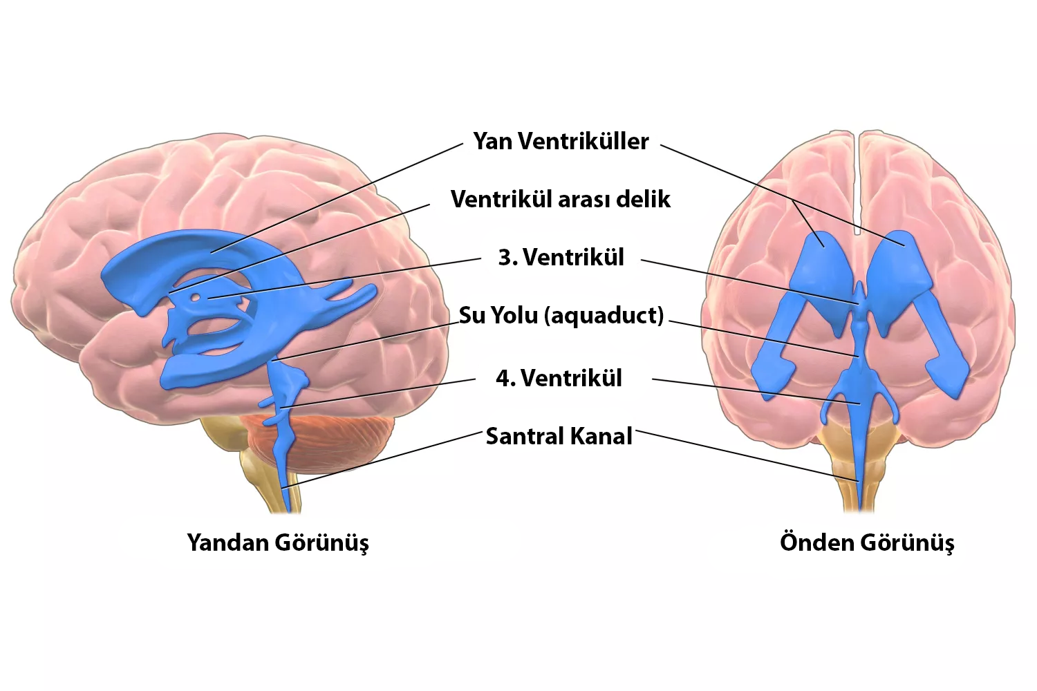
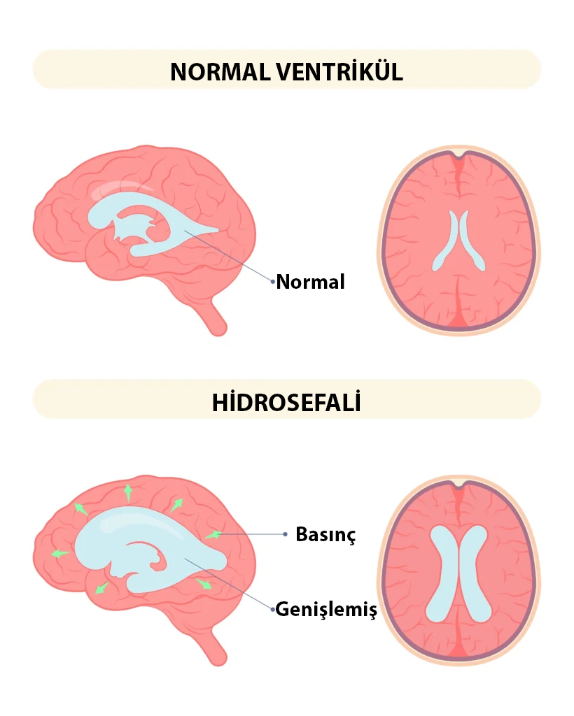
The brain contains fluid-filled cavities called ventricles. Cerebrospinal fluid (CSF) circulates through these spaces continuously and is produced and absorbed in a balanced way. Hydrocephalus develops when this balance is disrupted, most commonly due to obstruction of CSF flow or impaired absorption. As a result, excess CSF accumulates in the ventricles and increases pressure on surrounding brain tissue. In medical terminology, this condition is called HYDROCEPHALUS.
Babies with hydrocephalus may have the following findings:
- Excessive head growth in young children (macrocephaly)
- Nausea and vomiting
- Eyes looking downward - sunset eye appearance
- Restlessness - feeding difficulties
- Delays in developmental milestones
- Excessive sleepiness
- Seizures (epileptic attacks)
- In older children and adults, headache is most common
Why does hydrocephalus occur?
Any condition that disrupts CSF circulation can cause hydrocephalus. In some patients it begins before birth, while in others it develops later due to acquired neurologic disease.
The main causes of hydrocephalus are:
- Cerebral hemorrhage due to premature birth
- Brain hemorrhages due to head trauma
- Infections affecting the brain and central nervous system
- Tumors that cause excessive production of cerebrospinal fluid
- Brain tumors blocking cerebrospinal fluid circulation pathways
- Some genetic diseases and syndromes
- Developmental anomalies, especially myelomeningocele
Is Hydrocephalus a Serious Disease?
When hydrocephalus develops rapidly, it can become life-threatening if not treated promptly.
Fortunately, emergency deterioration is not the most common course. Many cases progress gradually, allowing time for thorough evaluation and planned treatment. A prompt neurosurgical consultation is still essential.
Age affects clinical presentation. In infants, open skull sutures may allow head enlargement before severe pressure symptoms appear. In older children and adults, this compensatory mechanism is limited, so symptoms may present differently and require timely intervention.
How is hydrocephalus diagnosed?
The diagnosis of hydrocephalus starts with suspicion. In infants, head circumference monitoring is one of the most important tools; a head circumference that grows more than expected over the following months is an important warning sign. In adults, clinical findings and patient complaints usually alert the neurosurgeon to the possibility of hydrocephalus. Because this disease can occur for many different reasons, radiologic imaging is necessary to understand why it developed and how the brain is affected. In most cases, the preferred imaging method is magnetic resonance imaging (MRI).
Is There a Treatment for Hydrocephalus? What are the Options?
When hydrocephalus is diagnosed, there are usually few treatment options other than surgery. If an identifiable mass is blocking the flow of cerebrospinal fluid (CSF), removing that mass and restoring normal flow may resolve the hydrocephalus. However, in many cases the blockage cannot be removed, and surgery is needed to divert the fluid.
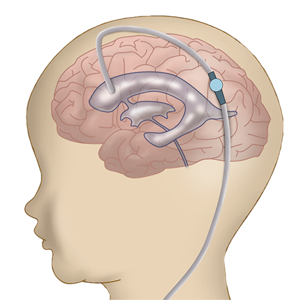
Shunt Systems
The main goal of hydrocephalus treatment is to redirect excess fluid accumulated in the brain to another part of the body, preventing it from compressing and damaging the brain. Although earlier attempts existed, shunt systems became the most effective solution after the 1950s. Medical devices called shunts carry excess fluid from the brain cavities to another body cavity through small tubes and valves, allowing it to be absorbed there. The most common destination is the abdominal cavity. Shunts can also be placed into the heart or the lining around the lung.
The most important advantage of shunt surgery is that it can be used for all types of hydrocephalus and can be performed in many hospitals because it is one of the first procedures neurosurgeons learn.
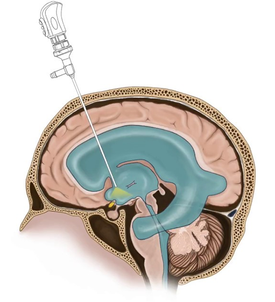
Endoscopic Third Ventriculostomy
In this relatively newer technique, an alternative pathway is opened for blocked CSF channels inside the brain. An endoscope with a camera is inserted into one of the brain chambers, and a small opening is made in the appropriate place so accumulated fluid can flow and be absorbed.
The most important feature of endoscopic third ventriculostomy is that it does not require placing a foreign device in the body. It aims to solve the problem through a more physiologic pathway inside the body.
Its most important disadvantage is that it is NOT SUITABLE FOR EVERY TYPE OF HYDROCEPHALUS. Not every case that can be treated with a shunt can be treated endoscopically. There are clear patient selection criteria. For example, it is not generally recommended for babies under 1 year of age, patients with a history of meningitis or brain hemorrhage, or patients who have already had shunt surgery. In these situations, the chance of success is much lower. It is most effective when MRI clearly shows that CSF pathways are blocked by a tumor or congenital anomaly. In such patients, success rates can reach 90%.
Another important issue is that it CANNOT be performed in every hospital or by every neurosurgeon. The procedure requires advanced and expensive surgical instruments, and the surgeon must have specific training in this area.
Shunt Surgery
Endoscopic Third Ventriculostomy
Are Hydrocephalus Surgeries Successful?
Before shunt systems took their modern form in the 1950s, hydrocephalus was a highly fatal disease. In medical history, very few treatments have affected human life as directly and clearly as shunt systems and shunt surgery.
However, it should not be forgotten that shunt surgery, like every surgical method, is not perfect and is not always one hundred percent successful.
Many factors, especially the patient's age and the underlying cause of hydrocephalus, affect the success of shunt surgery. The most important problem with shunts is that they can stop working. Shunts are made of very small tubes and valves. They can become blocked, break, or become infected. For this reason, patients with shunts may need more than one operation. This is a worldwide issue; patients treated in the United States, Japan or Turkey can all face it. The chance of shunt problems is highest in the first year and decreases over time.
This should never be interpreted as meaning that shunt surgery is an unsuccessful operation. SHUNTS SAVE LIVES. Every procedure has a possibility of problems; what matters is recognizing and managing those problems correctly.
Frequently Asked Questions
I should say from the start that there is no such thing as "easy surgery," and every type of surgery is important to us. On the other hand, shunt surgery and endoscopic surgery are relatively short and have a lower chance of problems compared with many other neurosurgical procedures. Still, as with any surgery, there are various complication risks, and your doctor will inform you about them.
Both types of surgery have advantages and disadvantages. In cases where both can be used, the endoscopic method is a more physiologic procedure and has a lower potential for problems than shunt surgery. However, it must be remembered that endoscopic treatment is NOT ALWAYS APPLICABLE.
Compared with many other neurosurgical procedures, these operations are not very large. The incisions are small and operation times are short. Children and adults usually tolerate these procedures very well.
Like other implanted medical devices, a shunt is used when it is medically necessary. In many patients, shunt systems support a stable daily life and long-term neurological health.