High-Risk Brain Tumors
For brain tumors that are deeply seated, located near critical areas, have been previously treated, or involve complex treatment decisions, a second opinion and treatment options may be considered.
Prof. Dr. Alp Özgün Börcek
Neurosurgery
Ankara Güven Hastanesi
You can schedule an appointment for a second consultation or examination.
Schedule an Appointment via WhatsApp Contact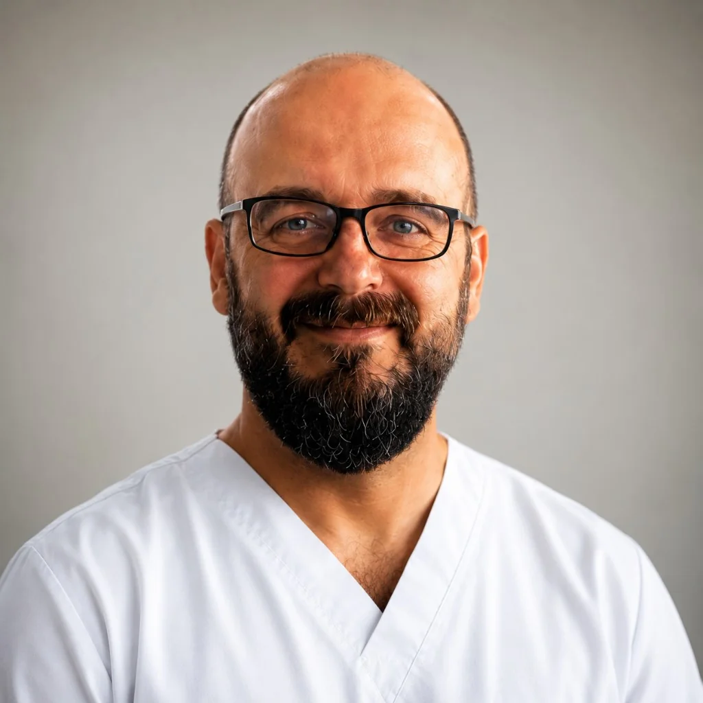
Prof. Dr. Alp Özgün Börcek
Neurosurgery
Ankara Güven Hastanesi
This content is for informational purposes only; a medical examination is required for diagnosis and treatment. Medical texts must be reviewed by a doctor.
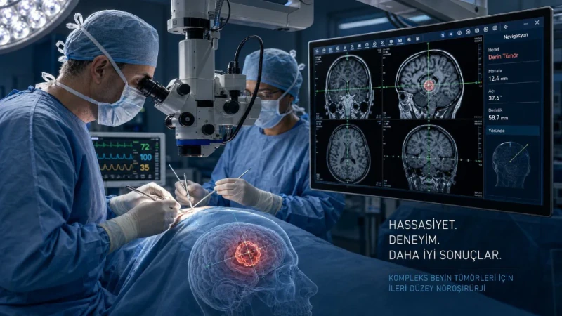
In some cases of brain tumor, the decision-making process is more complex: if the tumor is located near the speech, motor, or visual centers; if it is deeply seated; if the patient has previously undergone surgery or radiation therapy; or if there is a suspicion of recurrence (relapse). In everyday language, these cases are referred to as high-risk brain tumor It could be said that the “risk” here does not always mean malignant; rather, it indicates that the treatment plan must be carefully tailored to the individual patient. For general information, brain tumor You can check out our page.
Prof. Dr. Alp Özgün Börcek provides services at Ankara Güven Hastanesi for the evaluation of brain tumors that are deeply seated, located near critical regions, and have been previously treated, within the scope of pediatric and adult neurosurgery. This text is for informational purposes only; it does not constitute a guarantee of specific treatment. An examination is required for an individual diagnosis and treatment plan.
What Factors Increase the Risk of a brain tumor?
Two lesions of the same size may require very different approaches depending on their location and history of previous treatment. The following factors complicate the evaluation:
- Proximity to a critical area: Speech, movement, vision, sensation, or the area around the brainstem. Aggressive surgery in these areas may increase the risk of permanent neurological impairment.
- Deep settlement: The thalamus, basal ganglia, or similar deep structures. The risk associated with conventional open surgery may be high; a biopsy or radiosurgery may be the preferred option.
- Previous treatment: Previous surgery, radiation therapy, or chemotherapy. Tissue adhesions, wound healing, and the radiation field affect treatment planning.
- Recurrence: Suspected tumor recurrence or a new lesion. It is difficult to make a decision without knowing the previous pathology and treatments.
- Rapid growth or neurological impairment: The progression of symptoms increases the urgency; follow-up may not be appropriate.
- Unspecified diagnosis: There is a lesion on the MRI, but its type is unclear. In this case, a tissue diagnosis via stereotactic biopsy may be necessary.
Just because it’s high-risk doesn’t necessarily mean surgery is required. Sometimes, follow-up, a stereotactic biopsy, or Gamma Knife may be more appropriate. In these cases, an experienced evaluation and, when necessary, a multidisciplinary decision-making process are important.
What Do "Deep Settlement" and "Critical Areas" Mean?
Critical (eloquent) regions, are areas that control vital functions such as speech, arm and leg movement, vision, or balance. When treating tumors near these areas, the goal is often to proceed “as safely as possible”: it may not always be possible or appropriate to remove the tumor completely.
Deep-seated Access to tumors (such as those near the thalamus or brainstem) via open surgery is difficult. Commonly discussed options in these cases include:
- Stereotactic biopsy: Establishing a pathological diagnosis based on a small sample
- Elective surgery: If debulking (tumor reduction) is possible within safe limits
- radiosurgery (Gamma Knife): If the size and type are appropriate
- Cancer treatment + follow-up: Once the diagnosis has been confirmed
Categorical statements such as “No surgery” or “Surgery is absolutely necessary” do not apply to every case. The decision is made based on imaging findings, symptoms, the patient’s overall condition, and, if possible, pathology results.
What Changes in Previously Treated or Recurrent Tumors?
Suspected recurrence or a history of previous surgery or radiation therapy may require a complete revision of the treatment plan. Scar tissue, the previous radiation field, and existing neurological deficits can increase the risks associated with a new surgical procedure. Therefore, the following documents are particularly critical:
- Initial and follow-up MRIs (comparison of growth rates)
- Pathology report and, if available, molecular markers
- Surgical discharge summary and radiation therapy plan / dose information
- Oncology Follow-Up Notes
In some cases of recurrence, repeat surgery may be appropriate; in others, Gamma Knife, chemotherapy, or a supportive approach is prioritized. The goal is not to “repeat” the previous treatment, but to determine the most appropriate and safest course of action for the current situation. For an overview, brain tumor treatment options You can also check out that page.
When Is a Stereotactic Biopsy Considered?
Stereotactic biopsy allows for the collection of a small tissue sample for diagnostic purposes in cases of deep-seated or critically located lesions. The goal is not to remove the tumor, but to determine its type. It is particularly indicated in the following situations:
- There is a lesion on the MRI, but it is unclear whether it is benign or malignant
- open surgery appears to carry a high risk, but a treatment plan cannot be developed without a diagnosis
- If pathological confirmation is required for oncological treatment (chemotherapy/radiotherapy)
A biopsy also carries risks; however, in selected cases, it provides a diagnosis with a lower risk than open surgery. The biopsy result, subsequent Gamma Knife, guides the decision regarding surgical or medical treatment.
What Documents Are Required for the Application?
In high-risk cases, a current report alone is not sufficient; having a complete set of imaging series and previous treatment records directly impacts the quality of the decision. If possible, please bring the following or share them digitally before your appointment:
- Current brain MRI or CT scans (CD, USB, or digital link — a report alone is not sufficient)
- Previous views (for comparison and growth rate)
- Pathology Report (if available; including molecular results)
- Surgical and Treatment Discharge Summaries
- History and Plans for Radiation Therapy / Chemotherapy (dose, area, dates)
- The one you use list of medications (especially blood thinners)
Documents can be reviewed in advance; coming prepared for the examination appointment speeds up the process. In most cases, a neurological examination is also part of the decision-making process.
What Is Assessed During the Examination?
When assessing high-risk brain tumor cases, the MRI report is not the only factor considered. A comprehensive evaluation includes the following aspects (general framework: brain tumor Ankara):
- Symptoms: Headache, seizure, loss of strength, changes in speech or vision, loss of balance
- Neurological examination: Strength, senses, reflexes, coordination
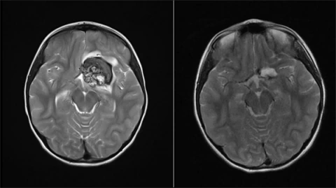
- Viewing: Location, size, edema, borders, comparison with previous MRIs
- Previous treatments: Surgery, radiation therapy, chemotherapy, and their side effects
- Pathology (if any): Type, degree, molecular properties
- General situation: Age, comorbidities, medications
The same MRI finding requires different approaches in different clinical presentations. Although an asymptomatic deep lesion and rapidly progressive neurological deficit may both be classified as “high-risk,” their levels of urgency differ.
What Options Are Discussed During the Decision-Making Process?
In high-risk cases, there is no single “right” approach; the options must be discussed openly:
- Surgery: Tumor reduction or resection using microsurgery and neuronavigation. The goal is to achieve the best possible tumor control within safe margins.
- Stereotactic biopsy: Low-risk sampling for diagnosis of deep or critical lesions.
- Gamma Knife / radiosurgery: In selected cases of small or recurrent tumors; this depends on the size, type, and location. For details, Gamma Knife You can check out the page.
- Follow: Regular MRI follow-up for some asymptomatic, slow-growing lesions.
- Chemotherapy / radiation therapy: A joint plan with oncology and radiation oncology.
The risks and benefits of each option are discussed on a case-by-case basis with the patient. The decision is often multidisciplinary is provided; no guarantee of a definite result is given.
How Does the Evaluation Process Work?
- Appointments and document sharing: MRI images, reports, and previous treatment records are submitted.
- Preliminary review: The images and files are reviewed.
- Examination: Symptoms and neurological status are evaluated together.
- Sharing options: The risks associated with surgery, biopsy, radiosurgery, follow-up, or oncological treatment options are explained.
- Decision: It is up to you whether to continue with the current plan, update it, or seek additional input.
If the documents are ready, most evaluations are completed in a single appointment. If necessary, additional imaging or a second opinion from another specialist may be recommended.
Why Is a Second Opinion Important?
Experiences and approaches may vary among different centers when dealing with high-risk cases. A second opinion can support the current treatment plan; clarify the timing of surgery, the need for a biopsy, or alternatives such as radiosurgery; and help you understand the risks and expected outcomes.
The goal is not to delay the process, but to make an informed decision. For the documents, the process, and the general second opinion approach, brain tumor second opinion You can check out the page. For general information on brain tumor brain tumor Ankara That page is also helpful.
What Should You Do in an Emergency?
It's okay to wait for an appointment non-existent Findings:
- Rapidly progressing loss of strength
- Impaired consciousness or speech impairment
- New or worsening seizures
- Vision loss or double vision
- A severe headache unlike any you've ever experienced
In these cases, go directly to the emergency room. If the patient is stable, it is appropriate to schedule an outpatient evaluation appointment.
For deep-seated brain tumors, those located near critical areas, or recurrent cases, a second opinion and treatment options may be considered. brain tumor and Gamma Knife You can also explore the main treatment options on our pages.
Schedule an Appointment via WhatsApp ContactRelated Pages


Frequently Asked Questions
No. In some cases, stereotactic biopsy, follow-up, or radiosurgery may be more appropriate. Surgery is not the first or only option for every high-risk tumor.
A patient’s history of previous surgery, radiation therapy, and chemotherapy, along with the pathological type and current MRI findings, are evaluated together. Surgery, radiosurgery, or oncological treatment is planned on an individualized basis.
The centers for respiration, heart rate, consciousness, and movement are located near this area; surgical risk may increase depending on the location. The decision should be made by an experienced team following a multidisciplinary evaluation.
No. A diagnosis can be made using stereotactic biopsy in deep and critical areas; radiosurgery or follow-up may also be considered.
Neurosurgery, radiation oncology, and, when necessary, medical oncology work together to evaluate the case. This approach is standard for complex cases.
If the documents are ready, most evaluations can be completed in a single appointment. If there are urgent neurological symptoms, you should go to the emergency department first.
No. Eligibility depends on the tumor’s size, type, location, and previous treatments; it is not suitable for every high-risk case. For more information, Gamma Knife You can check out our page.
If possible, a pathological diagnosis clarifies the condition and helps shape the treatment plan. In unclear cases, a biopsy may be necessary first.
No. The location and difficulty of treatment are what define the term "high-risk"; it does not always indicate malignancy. Benign tumors can also be considered high-risk if they are located in critical areas.
Yes. MRI images and reports can be sent digitally in advance. You may need to come to Ankara for the examination.
Author: Prof. Dr. Alp Özgün Börcek
Last updated: July 12, 2026
This content is provided for informational purposes only; it is not intended to replace a personal diagnosis or treatment.